
Andor live cell confocal imaging platformintegrated with CSU-W1 Spinning Disk Field Scanning Confocal System, iXon Ultra EMCCD and temperature control system, allow users flexible and powerful systems for a wide range of imaging applications. By using a disk containing microlens arrays in combination with the Nipkow disk, rotated to scan the entire field of view at high speeds, thus, making it possible to view confocal fluorescent images in real-time through the eyepiece of the CSU head. As compared to conventional single point scanning, multi beam scanning by the CSU requires a significantly low level of light intensity per unit area, which results in significantly reduced photo bleaching and phototoxicity in live cells. And then EMCCD camera has many technical advantages such as high sensitivity, high resolution, and high readout speed, which further enhances the imaging capabilities of Andorra Live Cell Confocal Imaging Platform.

Manufacturer:
CSU-W1 Confocal Scanner Unit
https://www.microscope.healthcare.nikon.com/products/confocal-microscopes/csu-series/csu-w1
iXon EMCCD Cameras
https://andor.oxinst.com/products/ixon-emccd-cameras
Specifications:
Confocal/wide field wavelength range |
λex:400-800nm λem:420-850nm |
Confocal speed |
400fps |
Laser wavelength |
405 nm,488 nm,561 nm,640 nm |
TIRF Imaging parameters |
405 nm,488 nm,561 nm,640 nm;60×Objective lens |
Wide field input power |
2W maximum for combined wavelength |
Camera |
EMCCD electron multiplying charge coupled device |
Confocal pinhole |
25μ and 40μm |
Active synchronization |
Laser irradiation synchronized camera exposure to minimize phototoxicity and photobleaching |
Output Power |
500mW/1W |
Horizontal resolution |
Limited by diffraction within 19mm diagonal |
Eyepiece magnification |
10× |
Objective magnification |
10× 20× 40× 60×(TIRF) 100× |
Size |
31.2 × 30.7 × 19.3 (cm) |
Location:112 room
Responsible: Qinglong Qiao
Training required:Training given by Qinglong
Booking:http://samp.cas.cn/admin.jsp (CAS)
Contact to Qinglong (Others)
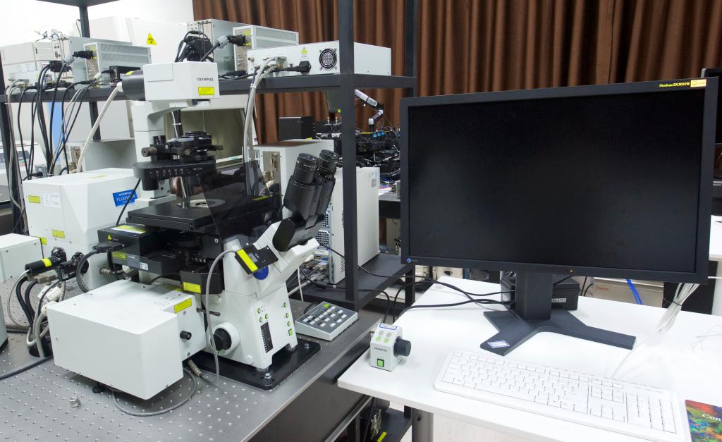
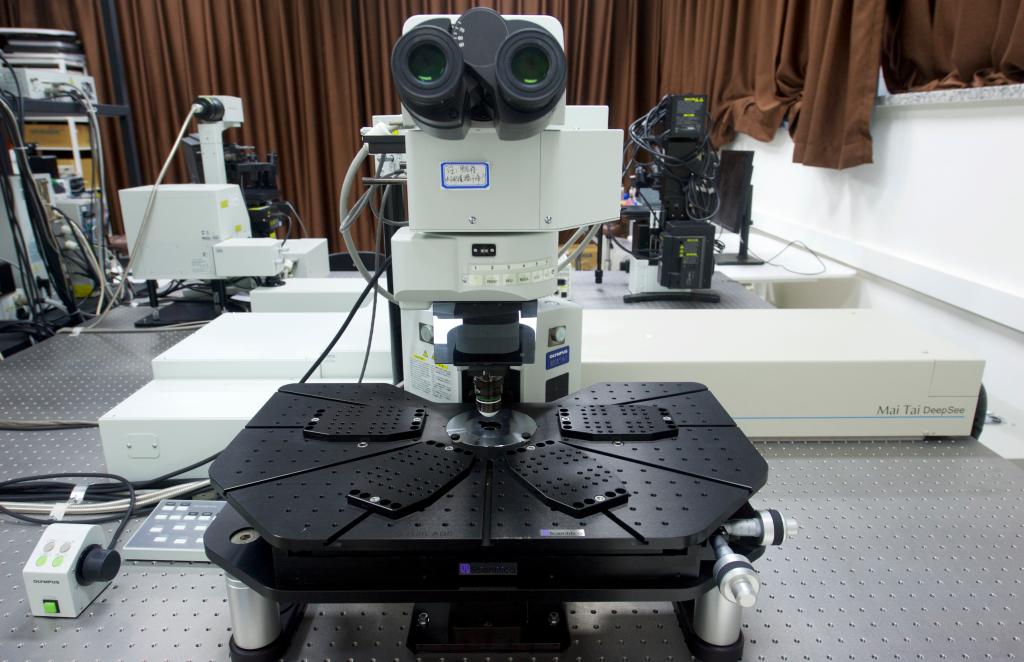
The Olympus FV1000 MPE confocal laser scanning microscopeoffers conventional confocal laser scanning of samples on slide. Supports multicolor fluorescent studies for imaging of living, whole mount or thickly sliced specimens. Dynamic biological processes can be imaged hundreds of micrometers within living cells and tissues. Low magnification lens and long working distance stage allow imaging of large samples. Provides support for applications where phototoxicity/photobleaching is a concern such as time course studies of living cells and tissues.
This microscope equipped with electric stage, CO2 heating incubation system, 5 lasers, 4 detection channels and 1 transmitted light detection channel. Single photon confocal microscope system was built on an Olympus inverted microscope IX81. Two-photon confocal microscope system was built on the Olympus research-grade upright microscope BX61WI.
Manufacturer:
https://www.olympus-lifescience.com.cn/zh/support/obsolete/
Specifications:
Single photon laser unit |
Laser: 405nm, 458nm, 488nm, 543nm, 635nm Motorized microscopes: Olympus IX81 |
Multi photon laser unit |
Mai Tai (Spectra-Physics) 690nm-1040nm Motorized microscopes: Olympus BX61WI |
Scanning method |
Light deflection via 2 galvanometer scanning mirrors |
Scanning speed |
(pixel time): 2 μs — 5ms |
Pixel size |
64×64 — 2048×2048 pixels |
High-speed scanning mode |
16 frames/sec (256×256) |
Eyepiece magnification |
10× |
Objective magnification |
5× 10× 20× 40× 60× 100× (IX81) 5× 10× 20× 25× 40× (BX61WI) |
Dimensions |
Time, Z, (wavelength) (or any combination thereof) |
Line scan |
straight line (includes rotation), free line, point XY scan |
Software |
FV10-ASW |
Required installation environment |
Room temperature: 20°C — 25°C ± 1°C, humidity: 60% or less, dust level: Class 10000, requires continuous (24-hour) power supply |
Power source environment |
AC100-120/220-240V 60VA |
Location:112 room
Responsible: Qinglong Qiao
Training required:Training given by Qinglong.
Booking:http://samp.cas.cn/admin.jsp (CAS)
Contact to Qinglong (Others)
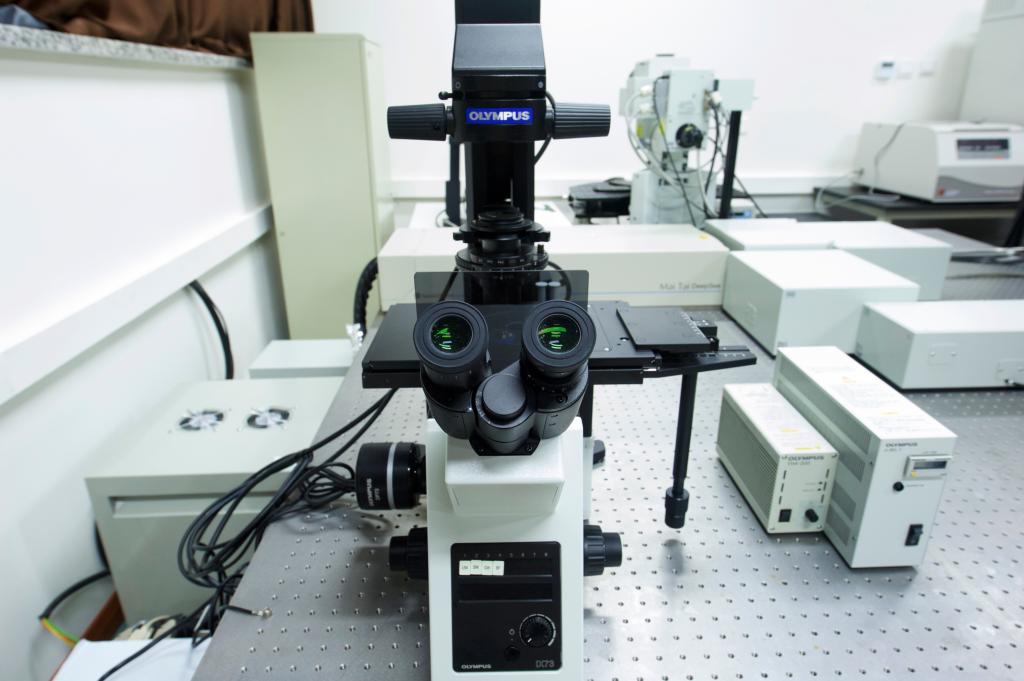
Olympus IX73 inverted microscopeis usually used for imaging living cells. It can perform fast fluorescence imaging, differential interference phase contrast imaging, phase contrast imaging and bright field imaging on cells according to different requirements of users. It has flexibly module configuration and high imaging resolution .
Manufacturer:
https://www.olympus-lifescience.com.cn/en/microscopes/inverted/ix73/
Maunal Book
https://www.manualslib.com/manual/662746/Olympus-Ix71.html?page=2#manual
Specifications:
Observation Method |
Fluorescence (Ultraviolet/Blue/Green/Excitation), Differential Interference Contrast (DIC), Phase Contrast, Brightfield |
Light source |
Mercury spectrum lamp |
Objective |
4×, 10×, 20×, 40× |
Optical system |
UIS2 |
Location:112 room
Responsible: Qinglong Qiao
Training required:Training given by Qinglong
Booking:Contact to Qinglong

© 2019 Dalian Institute of Chemical Physics, CAS
Molecular Probes & Fluorescence Imaging Group
All rights reserved. 辽ICP备05000861号
Professer
457 Zhongshan Road, Dalian. 116023, P. R. China
Phone: + 86- 041184379648
Emai: zcxu@dicp.ac.cn